| Western Pacific: off Japan, Taiwan and Australia. Collected from southeastern Japan (Okinawa trough, Tosa Bay, and Kyushu–Palau Ridge) at depth range 261–600 m. In Taiwanese waters this species is usually captured together with Lophiodes mutilus at depths of about 280–310 m, and some may range deeper. Specimens were collected from the Tasman Sea of southeastern Australia at depths of 300–750 m, and a series of photographs were taken of a specimen at a depth of 288 m at Greenwell Point, New South Wales, Australia, by an underwater vehicle (K. Aitken, personal communication, 20 March 2005). Two specimens were collected from the northwest shelf off Australia at depths of 492–520 m (Ref. 79601). |
|
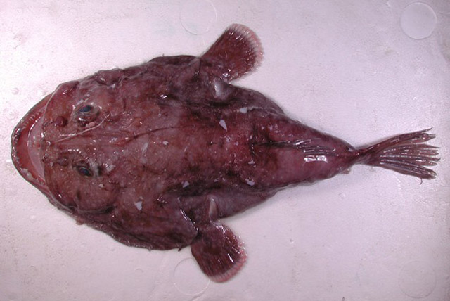
A species of the Lophiodes mutilus group differing from congeners by the following characters: a rounded esca with paler tip; third dorsal spine bearing a pair of black tendrils located at two-thirds of illicial length; 18–22 (mainly 20–21) pectoral fin rays; a relatively short head (33.4–39.6% SL); a relatively short illicium (16.5–26.7% SL); a relatively short third dorsal spine (30.0–42.3% SL); fifth dorsal spine relatively long, when folded back reaching base of third dorsal fin ray, and anterior frontal spine enlarged (Ref. 79601).
Tail cylindrical, somewhat depressed, tapering posteriorly; eye large; gill openings extending in front of pectoral fins; both maxilla and frontal ridge smooth, without knobs; frontal divided into three spines, anterior one enlarged, directed forward, sometimes divided into two sub-spines; sphenotic with two spines, inner spine well developed, straight and directed upward; inner frontal spine present in specimens smaller than 300 mm and reduced in specimens larger than 300 mm; single interopercular spine; parietal spine present at either side of third dorsal spine base, reduced in larger specimens; hyomandibula with two spines, anterior spine smaller, becoming blunt with three knobs in larger specimens; humeral spine well developed with three to four spines. All dorsal fin spines, except for the third, devoid of tendrils; second to sixth dorsal spines bearing a tiny dark bulb at tip in most specimens; illicium lightly pigmented, relatively short (16.5–26.7% SL), when folded back reaching third dorsal spine base, reaching sphenotic spines in larger specimens; esca a rounded bulb, slightly darker than illicium, tip pale; second dorsal spine slightly longer than illicium, when folded back reaching between third dorsal spine base and end of neurocranium; third dorsal spine relatively short (30.0–42.3% SL), bearing one pair of darkly pigmented tendrils at about two-thirds its length, when folded back reaches third dorsal fin ray base, reaching origin of soft dorsal fin in larger specimens; fourth dorsal spine absent; fifth dorsal spine relatively long, when folded back reaching base of third dorsal fin ray, reaching origin of soft dorsal fin in larger specimens; sixth dorsal spine very short, when folded back not reaching origin of dorsal fin, embedded under skin in larger specimens. Anal fin extending beyond caudal fin base in smaller specimens, reaching caudal fin base in larger specimens (Ref. 79601).
Coloration in life: based on a series of underwater photographs of a male specimen taken from Greenwell Point, New South Wales, Australia: dorsal surface pale brown with numerous medium-sized, diffuse light blue or gray patches; illicium paler; esca darker at base and paler at tip; some darker patches associated with head spines and upper jaw; pectoral fin margin pale, dorsal surface with some diffuse pale blue spots (Ref. 79601). |